
Our Health Library information does not replace the advice of a doctor. Please be advised that this information is made available to assist our patients to learn more about their health. Our providers may not see and/or treat all topics found herein.


Pelvic Ultrasound
Test Overview
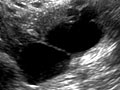
A pelvic ultrasound is a test that uses sound waves to make a picture of the organs and structures in the lower belly (pelvis).
This test looks at the bladder and:
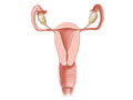
- The ovaries, uterus, cervix, and fallopian tubes of a woman (female organs).
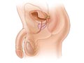
- The prostate gland and seminal vesicles of a man (male organs).
Organs and structures that are solid and uniform (such as the uterus, ovaries, or prostate gland) or that are fluid-filled (such as the bladder) show up clearly on a pelvic ultrasound. Bones may block other organs from being seen. Air-filled organs, such as the intestines, can make the image less clear.
Pelvic ultrasound can be done in several ways.
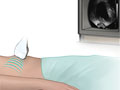
- Transabdominal ultrasound.
A small handheld device called a transducer is passed back and forth over the lower belly.
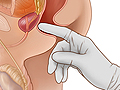
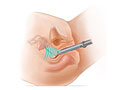
- Transrectal ultrasound.
The transducer is shaped to fit into the rectum. A transrectal ultrasound is the most common test to look at the male pelvic organs, such as the prostate and seminal vesicles. The test may also be done to look for rectal problems in men or women.
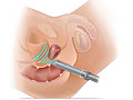
- Transvaginal ultrasound.
The transducer is shaped to fit into a woman's vagina. A woman may have both transabdominal and transvaginal ultrasounds to look at the whole pelvic area.
- Transperineal and translabial ultrasound.
- Both of these types of ultrasound are used on the outside of the genital area to look for urinary and pelvic problems.
In all of these ultrasounds, the transducer sends the reflected sound waves to a computer, which makes them into a picture that is shown on a video screen. Ultrasound pictures or videos may be saved as a permanent record.
Why It Is Done
For men and women, pelvic ultrasound may be done to:
- Find the cause of blood in the urine (hematuria). An ultrasound of the kidneys may also be done.
- Find the cause of urinary problems.
- Look at the size of the bladder before and after urination. This can determine whether the bladder is emptying completely during urination.
- Check for growths in the pelvis.
- Guide the placement of a needle during a biopsy or when draining the fluid from a cyst or abscess.
- Check for colorectal cancer and how it is responding to treatment.
Women
For women, pelvic ultrasound may be done to:
- Find out what is causing pelvic pain.
- Look for the cause of vaginal bleeding.
- Look for problems from pelvic inflammatory disease (PID).
- Find an intrauterine device (IUD).
- Look at the size and shape of the uterus and the thickness of the uterine lining (endometrium).
- Look at the size and shape of the ovaries.
- Check the condition and size of the ovaries during treatment for infertility.
- Confirm a pregnancy and see if it is in the uterus. Pelvic ultrasound may be used early in pregnancy to check the age of the pregnancy or to find a tubal pregnancy (ectopic pregnancy) or multiple pregnancy.
- Check the cervical length in a pregnant woman at risk for preterm labor.
- Check a lump found during a pelvic examination.
- Check uterine fibroids found during a pelvic examination. Pelvic ultrasound may also be done to check the growth of uterine fibroids.
- Guide a procedure to remove an ovarian follicle for in vitro fertilization.
Men
For men, pelvic ultrasound may be done to:
- Look at the seminal vesicles and the prostate gland.
- Check for prostate cancer. Other tests, including digital rectal examination, prostate-specific antigen blood test, and prostate biopsy, may also be used.
- See if urinary problems are being caused by a prostate that is getting bigger, such as from benign prostatic hypertrophy (BPH).
- Check to see if a problem with the prostate gland may be causing infertility.
How To Prepare
Wear loose clothes for the pelvic ultrasound. You may need to remove all your clothes below the waist and put on a gown before the test. If a man is also having a biopsy of the prostate gland, he may be given antibiotics for a day before the test.
How It Is Done
This test is done in an ultrasound room in a hospital, clinic, or doctor's office. If both a transabdominal and transvaginal ultrasound will be done, the transabdominal ultrasound will usually be done first.
You will need to remove any jewelry that might be in the way of the ultrasound. You will need to take off most of your clothes below the waist. You will be given a gown to use during the test.
You will lie on your back (or on your side) on a padded table.
You need to lie very still while the ultrasound is being done. You may be asked to take a breath and hold it for several seconds during the test.
Transabdominal ultrasound
For transabdominal ultrasound, you may need to drink 4 to 6 glasses of water about an hour before the test. Don't empty your bladder until the test is over. If you can't drink enough fluid, your bladder may be filled with water through a thin flexible tube (catheter) inserted into your bladder.
Gel will be put on your belly to improve the quality of the sound waves. A small, handheld device called a transducer is gently moved over your belly. A picture of the organs and blood vessels can be seen on a video screen.
When the test is done, the gel is cleaned off your skin.
Transrectal ultrasound
If you are having a transrectal ultrasound, you may need an enema before the test.
For transrectal ultrasound, you will be asked to lie on your left side with your knees bent. A digital rectal examination may be done before the ultrasound test. Then a lubricated transducer probe will be gently placed into your rectum. It will slowly be moved to take pictures from different angles. You may feel some pressure. Water may be put into your rectum to clean the end of the transducer so that clear pictures can be seen.
Transvaginal ultrasound
For transvaginal ultrasound, you will empty your bladder. You will be asked to lie on your back with your knees bent and feet and legs supported by footrests.
A thin, lubricated transducer probe will be gently placed into your vagina. It will slowly be moved to take pictures from different angles.
In rare cases, sterile saline is put in the uterus through a thin tube (catheter). This allows the doctor to look at the inside of the uterus (hysterosonogram).
How long the test takes
A pelvic ultrasound can take 15 to 30 minutes.
How It Feels
If you have a transabdominal ultrasound, you may feel pressure in your bladder and a strong urge to urinate because your bladder is full.
You will feel pressure from the transducer as it passes over your belly. If you have an injury or pelvic pain, the pressure of the transducer may be painful. You will not hear or feel the sound waves.
During a transvaginal or transrectal ultrasound, you will feel pressure from the transducer probe as it is put into your vagina or rectum.
Risks
There is little risk from a transabdominal or transvaginal ultrasound.
A transrectal ultrasound has a small risk for problems if a biopsy is done. Call your doctor if you have any problems after the test.
Results
Normal: | Your ovaries, cervix, and uterus have a normal shape and size and are in the normal place. No growths, tumors, fluid, or other problems, such as cysts, are present. Small cysts (follicles) in the ovaries of women who are able to have children are normal. |
|---|---|
If you are using an intrauterine device (IUD), it is in the uterus. | |
If you are pregnant, your baby (fetus) is developing inside the uterus. | |
Your bladder is normal in size and shape. No stones or abnormal growths are present. If the bladder is checked before and after urination, it empties completely. Urine flows normally from the ureters into the bladder. | |
If you are pregnant, a normal cervix measures long. | |
Abnormal: | Your uterus is big or abnormally shaped because of uterine fibroids. Cysts or tumors are present, such as cancerous or noncancerous tumors of the ovaries, uterus, or cervix. |
The thickness of the lining of the uterus (endometrium), called the endometrial stripe, is greater than normal. In some age groups, a thicker endometrial stripe (also called endometrial hyperplasia) may mean a higher chance of endometrial cancer. | |
Problems from pelvic inflammatory disease (PID), abscesses, kidney stones, or other problems are present. | |
An ectopic pregnancy is present. | |
An abnormal amount of fluid is present in the pelvis. | |
The bladder has an abnormal shape or a thick wall. A growth or stone is seen in the bladder. If the bladder is checked before and after urination, it may not empty completely during urination. | |
If you are pregnant, an abnormal cervix measures short. |
Normal: | Your prostate gland and seminal vesicles are normal in size and shape. No growths, tumors, or other problems, such as cysts, are present. |
|---|---|
Your bladder is normal in size and shape. No stones or abnormal growths are present. If the bladder is checked before and after urination, it empties completely during urination. Urine flows normally from the ureters into the bladder. | |
Abnormal: | Your prostate gland is enlarged (benign prostatic hypertrophy, or BPH). This is one of the most common abnormal findings. An abscess, a kidney stone in the urinary tract, or a tumor in or near the prostate gland or bladder may be present. |
The bladder has an abnormal shape or a thick wall. A growth or stone is seen in the bladder. If the bladder is checked before and after urination, it may not empty completely during urination. | |
An abnormal amount of fluid is present in the pelvis. |
Related Information
Credits
Current as of: July 26, 2023
Author: Healthwise Staff
Clinical Review Board
All Healthwise education is reviewed by a team that includes physicians, nurses, advanced practitioners, registered dieticians, and other healthcare professionals.
Current as of: July 26, 2023
Author: Healthwise Staff
Clinical Review Board
All Healthwise education is reviewed by a team that includes physicians, nurses, advanced practitioners, registered dieticians, and other healthcare professionals.


This information does not replace the advice of a doctor. Healthwise, Incorporated disclaims any warranty or liability for your use of this information. Your use of this information means that you agree to the Terms of Use and Privacy Policy. Learn how we develop our content.
To learn more about Healthwise, visit Healthwise.org.
© 1995-2024 Healthwise, Incorporated. Healthwise, Healthwise for every health decision, and the Healthwise logo are trademarks of Healthwise, Incorporated.